A fin de entender cómo funciona la resonancia magnética funcional (fMRI) es importante tener conocimientos básicos sobre el funcionamiento y la anatomía del cerebro. También es necesario conocer datos básicos sobre la imagen de resonancia magnética. Comencemos desde el principio:
Una curso muy corto en Neuroanatomía
El cerebro está dividido en dos hemisferios, el derecho y el izquierdo. Cada hemisferio se divide en lóbulos. Cada lóbulo tiene diferentes áreas como muestra la figura siguiente tomanda con autorización de
http://www/umich./edu%20cogneuro/Brodmann.html.
Cada región en un color diferente es un lóbulo. El amarillo es el lóbulo frontal; el verde el lóbulo parietal; el lóbulo occipital es el azul, mientras que el lóbulo temporal es rojo.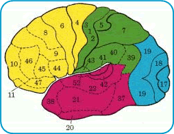
Las áreas 1, 2, 3 y 5 "sienten" las sensaciones del tacto, posición y temperatura. Las áreas 4, 6 y 8 mueven los músculos del cuerpo. Areas como las 44, 45 y 47 producen el lenguaje. Areas 41 y 42 son las áreas para "oír" y las 17 y 19 para "ver"
Otras áreas como la 37, 46, 10, 22, 38 están a cargo del proceso complejo de la memoria, lenguaje interno, comprensión y planeamiento.
Esta es una regionalización de la función. También hay algo llamado "lateralización" de la función. Eso significa que una actividad en particular puede estar predominantemente localizada en un hemisferio. Eso sucede con el lenguaje. En caso todas las personas diestras, el hemisferio izquierdo domina el lenguaje.
Cada cosa que hacemos, cada pensamiento, sensación o tacto es producida por la actividad de las células localizadas en un área específica del cerebro. Por ejemplo, si recibe la cuenta de su tarjeta de crédico y se aterra, es porque un grupo de células en la parte interior del cerebro comienzan a disparar, aumentando la corriente eléctrica, transmisión y el metabolismo. Estas células necesitan más sangre porque necesitan más combustible. Y sucede que cuando un área del cerebro se activa, esa área recibe más sangre porque en esa región hay dilatación de las pequeñas venas sanguíneas.
Por lo tanto, cualquier acción del cerebro causa un aumento de sangre en el punto de la corteza que rige la acción.
Ahora, sigamos a la segunda parte.
Un curso muy, muy corto en Imagenes de Resonancia Magnética (MRI)
La imagen de resonancia magnética es un procedimiento para ver la anatomía de los órganos internos del cuerpo. Se basa en tres cosas: magnetismo, radiofrecuencia y computadoras.
A fin de entender cómo estas tres cosas no muestran la anatomía del cerebro (y del cuerpo entero) es importante saber cinco otras cosas:
- Nuestro cuerpo contiene grandes cantidades de agua.
- El agua esta compuesta de hidrógeno y oxígeno (el hidrógeno se presenta básicamente como protones).
- Los protones son cargas positivas que giran.
- La carga que gira crea un campo magnético que puede orientarse. En otras palabras, estos micromagnetos pueden ir hacia arriba, hacia abajo, derecha o izquierda.
- La giración de estos micromagnetos tiene una frecuencia (velocidad) específica.
Y eso es todo. Regresemos a las tres cosas necesarias para realizar una imagen de resonancia magnética (MRI): magnetismo, radiofrecuencia y computadoras.
Magnetismo
La máquina de resonancia magnética es un imán grande y potente. Cuando el cuerpo está dentro de la máquina, cada protón del cuerpo se orienta de la misma manera (por ejemplo, con el poste positivo hacia arriba). Eso significa que el cuerpo se convierte en un imán; pero no un imán hemógeno, porque la cantidad de agua en cada parte del cuerpo varía de acuerdo con las características específicas del órgano: la capa, el lugar y hasta los tipos de células. Por lo tanto, el cuerpo humano puede retratarse como un mapa tridimensional de cambios en el campo magnético con la forma de un cuerpo humano. Si conocemos la densidad de eos cambios podemos obtener imágenes de la anatomía interna. ¿Y como sabemos la densidad? Por medio de la radiofrecuencia.
Radiofrecuencia
La radiofrecuencia da la energia que hace girar los protones, aumentando la amplitud de sus giros sin cambiar la frecuencia. Ahora el mapa magnético tridimensional se convierte en un mapa tridimensional de energía. Cada punto específico del cuerpo tiene una energía particular (o intensidad, en términos de radiofrecuencia). Al dejar de aplicar la radiofrecuencia al cuperpo, los giros de los protones vuelven a su estado original y en ese momento liberan ondas de radiofrecuencia. Ahora tenemos un mapa tridimensional de radiofrecuencia. Y la radiofrecuencia puede ser registrada con espirales.
Computadoras
El resto del procedimiento se hace por computadoras que convierten la intensidad de la señal, la fase de la señal y la localidad de la señal en una matriz de puntos con diferentes valores. Cada valor está representado con un tono de gris. El valor mínimo es negro, y el valor máximo es blanco, y en el medio hay una escala de grises. Ahora tenemos LA IMAGEN DE RESONANCIA MAGNETICA
Cómo funciona la Imagen de Resonancia Magnetica Funcional
En la imagen funcional tenemos dos componentes: una tarea y un resultado. La tarea es la acción o actividad que el sujeto ejecuta a fin de producir una activación específica en el cerebro. Por ejemplo, mover los dedos de la mano derecha conínuamente es una tarea motora que "activa" la corteza cerebral en el lóbulo frontal izquierdo. El resultado en la imagen de resonancia magnética funcional es una imagen de muestra esta activación.
La tarea puede ser de cualquier tipo. Motora, sentir el tacto, tener una percepción, pensar en palabras abstractas, responder a un estímulo cambiante, escuchar música, comprender una historia y muchas otras. La tarea produce la siguiente secuencia de eventos:
- Aumenta el metabolismo en el área del cerebro involucrada en la tarea.
- Aumenta el volumen de sangre en esa región específica.
- Aumenta el nivel de oxígeno.
- Cambios en el campo magnético local (Se recuerda del mapa de campo magnético tridimensional que discutimos anteriormente?)
- Cambios en la intensidad de la energía (radiofrecuencia) en la misma área.
En breve, la tarea evoca ctividad en una región del cerebro, y esta actividad cambia la intensidad de la radiofrecuencia que sale de esa parte. Este es un cambio muy pequeño. Pero, se repetimos la tarea varias veces podemos sumar los cambios hasta llegar a un resultado significante que puede ser registrado.
Comparamos la actividad del cerebro de una persona durante una tarea en particular con el nivel de actividad mientras descansan. Sin embargo, el cerebro funciona contínuamente, aún cuando se descansa. Por esa razón, el nivel durante el descanso se usa como línea básica de la actividad fundamental del cerebro.
Es necesario que sepamos el valor de la señal de actividad fundamental y el valor de la señal relacionada con la tarea. Para hacerlo, por unos minutos tomamos cientos de imágenes de la región blanco. Mientras tanto, el sujeto alterna entre períodos de actividad (realizando la tarea) y períodos de descanso. De esta manera tenemos un grupo de imágenes de una región tomadas durante la tarea, y el número de imágenes de la misma regiónb tomadas durante el descanso. Se obtiene un promedio de dos imágenes correspondiendo a dos condiciones en la cual una es "activada".
Como hemos visto anteriormente, las imágenes son el resultado de los valores de la intensidad de la señal, codificadas en la escala gris. Estos valores pueden sumarse, restarse, etc. Ese es el próximo paso. Computadoras de gran potencia restan los valores de línea básica de las activadas. Estos valores de activación son transormados en un mapa a colores. Usualmente la escala de este mapa varía de azul a rojo en forma creciente. Finalmente, los colores son superimpuestos sobre imágenes anatómicas, de una manera similar a los mapas del tiempo que se superimponen a ilustraciones geográficas.
Cómo la imagen de resonancia magnética funcional ayuda en la Medicina
La imagen de resonancia magnética funcional tiene una terminologia particular. Las siguientes imágenes explican los términos más importantes que se usan para describir un experimento.
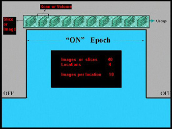
Un período es el tiempo en que ocurre algo.
ON: El nombre del período en el cual se realiza la tarea
OFF: El nombre del período en el cual el sujeto descansa
Un escán es la imagen obtenida en cada unidad de tiempo.
Un nivel es la localización del sitio a explorar. Se pueden explorar varios niveles al mismo tiempo.
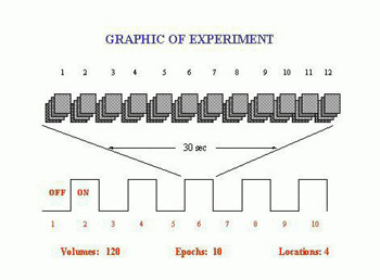
En este gráfico se muestran: 5 períodos ON, durante el cual el sujeto realiza la tarea; 5 períodos OFF, durante el cual el sujeto descansa; cada período tiene 30 segundos de duración por una cantidad total de 5 minutos de duración del experimento. Se exploran cuatro niveles del cerebro, con 120 imágenes (o escanes) divididos alternamente en grupos de 12.
Un resultado final
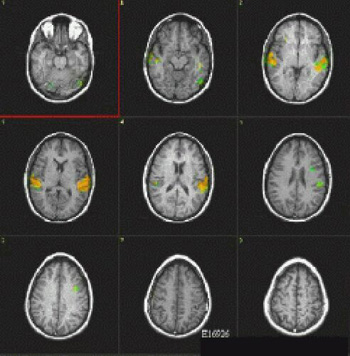
Las áreas a color muestran la activación de las regiones del cerebro involucradas en oír y entender la voz humana. El hemisferio izquierdo muestra una activación mayor.